Pushing the Boundaries of Human Brain Imaging
A next-generation fMRI machine, the centerpiece of the new MindCORE Neuroimaging Facility, gives researchers across campus a novel tool to study the mind-brain connection.

To study the human brain, really study it, and its relationship to the human mind, relatively little compares to using functional magnetic resonance imaging (fMRI), according to Joseph W. Kable, Jean-Marie Kneeley President’s Distinguished Professor of Psychology & Marketing.
“It allows you to study the entire brain at the same time, know where the signals are that you’re recording from, and do it with decent spatial resolution,” says Kable, who is also director of MindCORE, Penn’s hub for the integrative study of the mind. “fMRI is a work-horse technology for trying to understand how the brain creates the mind, the relationship between the human brain and behavior.”
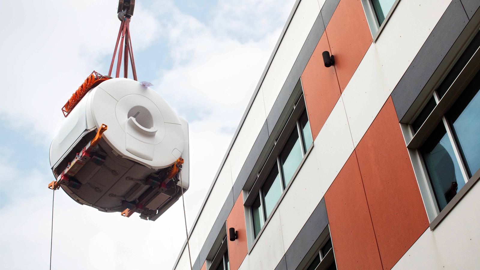
Now MindCORE has in its stable of tools its very own fMRI, lowered by crane into the Pennovation Lab building at Pennovation Works in mid-April. It’s the centerpiece of the new MindCORE Neuroimaging Facility established under the Mapping the Mind goal outlined in the Penn Arts & Sciences strategic plan.

According to Joseph W. Kable, director of MindCORE, the goal of the new fMRI machine is to provide access to the resource for a large community of people across the University.
The machine, a MAGNETOM Cima.X 3 Tesla magnetic resonance imaging whole-body scanner, received approval from the Food and Drug Administration in early 2024. It’s the newest fMRI iteration from the company Siemens Healthineers and offers incredibly high-resolution images quickly, with a unique GEMINI gradient coil system that makes visible previously hidden underlying structures.
It was up and running at the Pennovation Lab in May, and Kable says he hopes data collection can begin as early as June. “The goal here is to provide access to the resource for a large community of people” across the University, he says.
Bringing the magnet to MindCORE has been several years in the making. Excited conversations began in 2019, but then the pandemic, coupled with the desire to wait for the “next generation” of fMRI to become available, stalled the plans. Once FDA clearance came through, the installation itself became its own undertaking, says Andrew Jones, Engineering Projects Manager in the School of Arts & Sciences’ Facilities Planning & Operations.
“The space to hold the fMRI is underground, so we dug a pit outside of the building. It’s a concrete vault,” explains Jones, who oversaw the project. “Because these machines are so heavy, you often put them on the ground floor of whatever building they’re in. Sometimes you have to get creative in how to get them there.”
In this case, that meant using a crane to lift and lower the fMRI into the room—one where the floor had been isolated from everything around it to prevent vibrations from affecting the machine’s functionality—then sealing the space from above. Should the machine need future servicing or updating, the “lid” should allow for easier access, Jones says. All told, the install took three weeks.
Though Penn isn’t the only university with such a machine strictly for this kind of research, it’s one of just a few. “The time had come for Penn to have such an important method for basic research, a facility dedicated to basic research on the human brain,” says Kable, who envisions using the machine for his own work, which focuses on decision-making.
Our researchers are eager to use this tool to push the boundaries of human brain imaging, to better understand the relationship between the mind and brain.
“One project we’ll probably start with is looking at individual differences in selfishness versus altruism and the hypothesis that people who are more altruistic have a greater affective empathy toward others,” he says. “There’s a set of brain regions that have been identified as responding when people see others in distress. It’s the same ones that respond when you yourself are in distress. We’re interested in showing that those responses predict who will be more giving or altruistic of others.”
Because the work of many MindCORE researchers includes children, there are plans to purchase a mock MRI that these youngest of study subjects can use to get acquainted with before going into an MRI machine. MindCORE also hired an executive director, Brock Kirwan, who began May 1. “Our researchers are eager to use this tool to push the boundaries of human brain imaging,” Kable says, “to better understand the relationship between the mind and brain.”


